This demo is for research & educational use only — not a medical device. Read full disclaimer.

AI-GS: Experimental AI-Based Glaucoma Risk Screening

(Non-Diagnostic Research Demonstration)
This tool is a research prototype for demonstration purposes only. It is not a medical device and does not provide diagnosis, treatment, or clinical decision support. Always consult a qualified ophthalmologist.
Uploaded images may be used, without personal identifiers and in aggregated form, to help improve the system over time. Please upload only images that you are permitted to use.
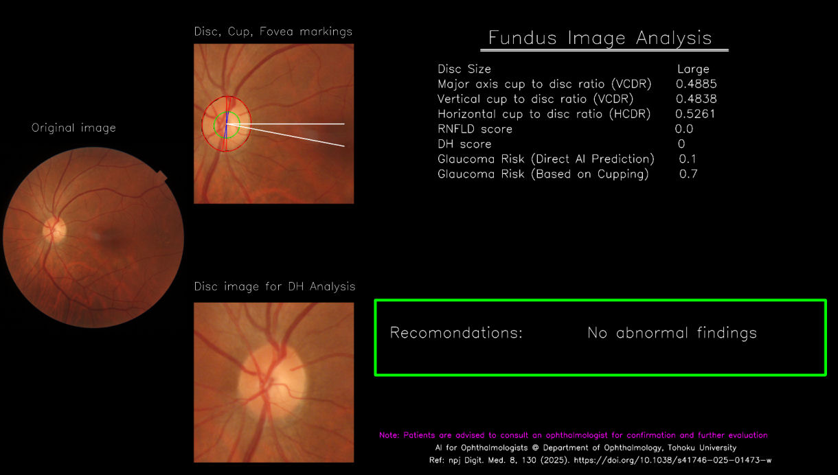
Upload a fundus image centered on the macula (fovea)
Images focused mainly on the optic disc or images that are blurry may not be processed correctly, as the model detects both the fovea and optic disc to estimate optic disc size.
0.0s
Example Input
Example macula-centered fundus image

Upload a macula-centered (fovea-centered) color fundus image.

  • Optic disc and macula should be visible
  • Image should be well-focused
  • Disc-centered/blurred images may fail/produce incorrect results

The system detects both the fovea and optic disc to estimate anatomical relationships.

Example Output
Example AIGS screening output

The output visualization provides:

  • Annotations of key anatomical structures, optic disc, optic cup, and fovea
  • Measurements of vertical and horizontal cup-to-disc ratios (VCDR, HCDR)
  • Probabilities of glaucoma risk, and retinal nerve fiber layer defect (RNFLD),
  • Visual marking of DH and its probability, if detected
  • Screening recommendation summarizing overall risk profile

Results are provided for research screening only and are not diagnostic.